
Magnetic Resonance Imaging (MRI) is an advanced medical imaging procedure that captures detailed images of the brain and its surrounding nerve tissues. It allows specialists to view the brain’s structure from multiple angles and assess features that might not be apparent through other imaging methods. A brain MRI also enables physicians to examine brain anatomy and detect abnormalities. Here are some benefits of using MRI to diagnose neurological conditions:
Provides Detailed Imaging
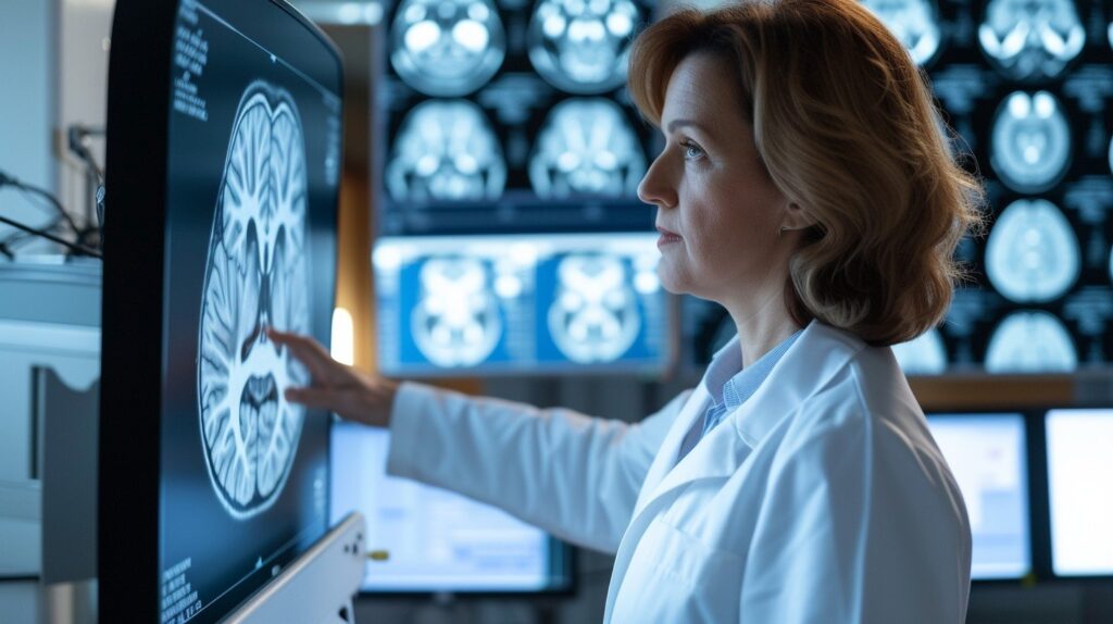
MRI scans produce high-resolution images of the brain’s soft tissues. This makes it possible to distinguish between gray matter, white matter, and cerebrospinal fluid. Radiologists often use MRI to examine the hippocampus, which impacts memory, or the brainstem, which controls basic body functions. The level of detail provided by MRI helps identify lesions, swelling, or malformations that might go unseen on an X-ray or CT scan.
MRI images are also three-dimensional. This allows doctors to view the brain from different perspectives and closely examine the regions that support speech, movement, and vision. It also reveals the precise localization of irregularities, guiding specialists as they develop a plan for the next steps in your care.
Healthcare teams often use MRI images to track changes in the brain over time. For patients with chronic neurological conditions, regular brain MRI scans help monitor progression and support decisions about ongoing management. By identifying even minor anatomical changes, doctors can adapt their recommendations and support overall patient well-being.
Detects Diverse Conditions
A brain MRI is a versatile tool in diagnosing a wide range of neurological conditions. Healthcare providers may request an MRI if a patient reports symptoms like persistent headaches, sudden weakness, unexplained seizures, or changes in cognitive abilities. An MRI can reveal areas of the brain affected by a recent stroke, helping doctors assess blood flow disturbance. MRIs can also:
- Detect brain tumors: Precise imaging helps differentiate between benign and malignant tumors. It can also show how a tumor affects surrounding structures.
- Monitor multiple sclerosis: An MRI highlights plaques or lesions in the white matter, which are characteristic of the disease’s progression.
- Evaluate trauma: A brain MRI assists in evaluating brain injuries sustained during trauma, including concussions or bleeding within the brain tissue.
- Assess development: In children, an MRI can assess developmental anomalies, such as those affecting the growth of certain brain regions.
Offers Non-Invasive Diagnosis
A brain MRI is a non-invasive procedure that requires no incisions or skin penetration. During the scan, you’ll lie on a motorized table that slides into the MRI machine’s opening. The equipment can make loud tapping or knocking noises, so patients are often given earplugs or headphones to help make the experience more comfortable. The process is painless and only requires you to stay still throughout the scan.
A contrast agent may be used to highlight certain blood vessels or tissue abnormalities. The contrast is administered through a small IV in your arm, and is generally well-tolerated. All imaging is conducted externally; there is no need for surgery, and no recovery time after the scan.
Schedule a Brain MRI Today
An MRI offers clear, detailed information, allowing your healthcare provider to thoroughly evaluate your neurological health. It non-invasively identifies a range of conditions, guiding your next steps in care. If you experience unexplained headaches, vision changes, or memory issues, a brain MRI could provide answers. Contact your doctor today to learn if an MRI scan is right for you.
- Expert Tips for Locating Quality Pinched Nerve Treatment Near You
- Understanding QuantumRF Technology In Skin Rejuvenation
- The Role of Regular Podiatry Check-ups for Overall Wellness
- Understanding the Role of a Plastic Surgeon in Cosmetic Procedures
- The Connection Between Shingles and Your Immune System