
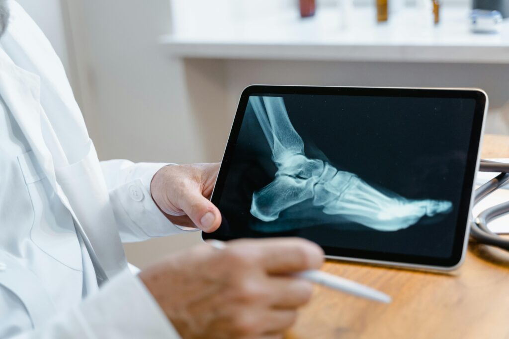
X-ray imaging has long been central to orthopedic care, providing a clear look at bones, joints, and surrounding structures. As technology advances, modern X-ray systems have become faster, more precise, and more adaptable to a variety of clinical settings. These changes have improved both the quality of diagnoses and the experience patients have during orthopedic evaluations.
Improving Image Quality with Digital X-Rays
In orthopedic care, imaging supports decisions about treatment for injuries, degenerative joint conditions, spine alignment, and congenital abnormalities. The evolution of X-ray tools has allowed providers to see structural issues in greater detail while exposing patients to lower doses of radiation. With better resolution and more flexible applications, today’s X-ray technology helps guide orthopedic care from initial consultation to post-treatment follow-up.
One of the most significant improvements in recent years is the shift from film-based to digital X-ray systems. Digital radiography uses sensors instead of film, which allows providers to capture and view images almost instantly. This quick turnaround can reduce wait times for patients and helps orthopedic specialists begin treatment planning sooner.
In addition to speed, digital X-rays offer sharper images. This improved clarity helps detect subtle changes in bone density, alignment, or healing progress. When providers can see more detail, they can make more accurate decisions about how to proceed, whether it involves physical therapy, bracing, or surgery.
Reducing Radiation Exposure for Patient Safety
Advances in equipment design and image processing have also led to reduced radiation exposure. While X-rays have always used relatively low doses, newer systems are calibrated to use the smallest amount necessary to achieve a diagnostic-quality image. For orthopedic patients who require multiple images—such as those with scoliosis, healing fractures, or joint replacements—this improvement offers a safer long-term experience. Some facilities now use dose-tracking software, which monitors exposure levels over time and alerts clinicians when cumulative doses approach certain thresholds. This helps orthopedic teams tailor care without compromising patient safety.
Enhancing Diagnostic Precision with 3D Imaging
While traditional X-rays provide a two-dimensional image, newer systems can generate 3D reconstructions from multiple views. This is especially useful in orthopedic care, where understanding the angles and spatial relationships between bones is often critical.
For example, in spine assessments, 3D X-ray images help visualize curvature and rotation that may not be as clear on a standard film. Similarly, in joint replacement planning, 3D models allow surgeons to select implants and plan procedures more precisely. These models can be used alongside CT or MRI scans for a complete picture of the musculoskeletal system.
Increasing Flexibility with Portable Systems
Orthopedic providers increasingly use portable digital X-ray units that can be brought directly to patients in hospitals, clinics, or post-surgical recovery rooms. These systems allow for imaging without transferring patients who may be in pain, immobilized, or recovering from surgery. This portability also supports care in outpatient settings, making follow-up visits more efficient. Images can be reviewed on-site, often within minutes, so that patients leave with a clear understanding of their next steps.
Modern X-ray systems are often integrated with electronic health records (EHRs), allowing images to be stored, reviewed, and shared securely with other members of a patient’s care team. For individuals receiving care across specialties—such as orthopedic surgeons, primary care physicians, and rehabilitation providers—this coordination helps support continuity and informed decision-making. Patients can also access their images through secure portals, which gives them more visibility into their care and helps them stay informed about their progress.
Explore More
As innovation continues, X-ray imaging in orthopedic care is becoming more tailored, efficient, and patient-centered. From digital clarity to lower radiation and improved mobility, these advancements offer both clinical and comfort-related benefits. For patients seeking orthopedic care, these improvements mean faster diagnoses, better outcomes, and a more streamlined experience across the course of treatment.
- Expert Tips for Locating Quality Pinched Nerve Treatment Near You
- Understanding QuantumRF Technology In Skin Rejuvenation
- The Role of Regular Podiatry Check-ups for Overall Wellness
- Understanding the Role of a Plastic Surgeon in Cosmetic Procedures
- The Connection Between Shingles and Your Immune System